 Same and next-day pediatric appointments
November is Diabetes Month, when we can take the opportunity to increase awareness of the struggles
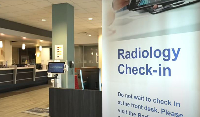 Optum Radiology at Crystal Run Healthcare
We are proud to share that Optum has made a significant investment in our Radiology services to improve patient care and access to leading-edge technology.